
Concussion risk in youth football
January 21, 2020
Science Daily/Virginia Tech
For decades, there's been a widespread assumption among people with an interest in sports-related injury that youth football players are more vulnerable to concussion and other head injuries than their older, bigger counterparts.
The Virginia Tech Helmet Lab has published the first data validating that conventional wisdom, in a study released in the January issue of the Annals of Biomedical Engineering.
The research, funded by a five-year grant from the National Institute of Neurological Disorders and Stroke of the National Institutes of Health, demonstrated that the head accelerations leading to concussion in youth football players are lower than those that typically cause injury in high school, collegiate, and professional athletes -- data that will be critical for designing and testing protective equipment.
"These are the first biomechanical data characterizing concussion risk in kids," said Steve Rowson, an associate professor of biomedical engineering and mechanics and the director of the Virginia Tech Helmet Lab. "Children aren't just scaled-down adults: Differences in anatomy and physiology, like head-neck proportions and brain development, contribute to differences in tolerance to head impact. These results can lead to data-driven interventions to reduce risk in youth sports."
Kids between the ages of 9 and 14 make up the largest group of football players in the country. But the research cataloging head impacts on U.S. football fields, and determining which ones are most likely to cause injury, has focused on high school, collegiate, and pro players, limiting the understanding of what conditions lead to concussion in youth football.
In younger players, the fatty myelin sheaths that help protect brain cells haven't fully developed. They also tend to have larger heads relative to their bodies than adult players do, with less neck musculature to help absorb the force of an impact. For all these reasons, researchers had presumed that youth players were more susceptible to concussion. But they needed data to prove it.
Virginia Tech has been studying head impacts in football since 2003, when Stefan Duma, the Harry Wyatt Professor of Engineering and founder of the Helmet Lab, instrumented the helmets worn by the university's varsity football team with sensors to measure head impacts.
Since then, Duma, Rowson, and their research team have expanded their studies to more sports and a broader range of demographic groups. They've been studying youth players since they won this critical grant from the NIH in 2015.
For the study, they partnered with researchers at Brown University and Wake Forest University to track six different youth football teams in Virginia, North Carolina, and Rhode Island. More than 100 players wore helmets lined with sensors that measured the linear and rotational acceleration of their heads during four seasons' worth of practices and games, recording thousands of impacts. All concussions were diagnosed by clinicians at each site; neuropsychological testing before and after each season measured the players' cognitive function.
The National Operating Committee on Standards for Athletic Equipment, an independent standards body that certifies equipment including football helmets, provided additional funding that helped accelerate the data collection.
That data allowed the researchers to develop a mathematical relationship between the magnitude of a head impact and the risk of concussion. What they found was that youth players are on average more susceptible to concussion at lower levels of acceleration than high school and collegiate players are.
In high school and college players, the average concussive impact is associated with a head acceleration around 102 g, with similar values for pro athletes. In youth players, the new study found, the average concussive impact was associated with a head acceleration of only 62 g. The rotational acceleration values associated with concussion were similarly reduced, from 4,412 rad/s2 in adults to 2,609 rad/s2 in youth players.
"These numbers prove for the first time that youth players are at a higher risk of injury at lower head accelerations," Duma said, "but it is important to note that the overall head acceleration exposure in youth football is much lower than in adult football."
Despite that heightened susceptibility, concussions in youth football are relatively rare: Younger, lighter players collide with less force than adult athletes, so they're less likely to jostle their brains enough to cause serious injury.
But as with any sport, there's still a risk of injury. So effective protective equipment is critical -- and that's the practical value of this study, Duma explained. Knowing the levels of acceleration that put youth players at risk for concussion provides a benchmark to use for helmet testing.
Earlier this year, the Helmet Lab released the industry's first youth-specific football helmet ratings, evaluating the helmets with laboratory tests based on their on-field data.
"No one had ever come up with a rating system tailored to youth helmets, partly because the data didn't exist," said Duma, who also directs the university's Institute for Critical Technology and Applied Science. "Now we can evaluate helmets based on the actual risks youth players experience, and companies can use that information to design models specifically for this large group of players."
https://www.sciencedaily.com/releases/2020/01/200121133259.htm
Blue light can help heal mild traumatic brain injury
January 15, 2020
Science Daily/University of Arizona
Daily exposure to blue wavelength light each morning helps to re-entrain the circadian rhythm so that people get better, more regular sleep which was translated into improvements in cognitive function, reduced daytime sleepiness and actual brain repair.
Early morning blue light exposure therapy can aid the healing process of people impact by mild traumatic brain injury, according to new research from the University of Arizona.
"Daily exposure to blue wavelength light each morning helps to re-entrain the circadian rhythm so that people get better, more regular sleep. This is likely true for everybody, but we recently demonstrated it in people recovering from mild traumatic brain injury, or mTBI. That improvement in sleep was translated into improvements in cognitive function, reduced daytime sleepiness and actual brain repair," said William D. "Scott" Killgore, psychiatry professor in the College of Medicine -- Tucson and lead author on a new study published in the journal Neurobiology of Disease.
Mild traumatic brain injuries, or concussions, are often the result of falls, fights, car accidents and sports participation. Among other threats, military personnel can also experience mTBI from exposure to explosive blasts: Shockwaves strike the soft tissue of the gut and push a burst of pressure into the brain, causing microscopic damage to blood vessels and brain tissue, Killgore said.
"Your brain is about the consistency of thick Jell-O," he said. "Imagine a bowl of Jell-O getting hit from a punch or slamming against the steering wheel in a car accident. What's it doing? It's absorbing that shock and bouncing around. During that impact, microscopic brain cells thinner than a strand of hair can easily stretch and tear and rip from the force."
Those with a concussion or mTBI might can momentarily seen stars, become disoriented, or even briefly lost consciousness following the injury; however, loss of consciousness doesn't always happen and many people who sustain a concussion are able to walk it off without realizing they have a mild brain injury, according to Killgore. Headaches, attention problems and mental fogginess are commonly reported after head injuries and can persist for weeks or months for some people.
Few, if any, effective treatments for mTBI exist. The U.S. Army Medical Research and Development Command funded the research to find alternatives to medicinal methods of mTBI recovery.
"About 50% of people with mTBI also complain that they have sleep problems after an injury," Killgore said.
Recent research has shown that the brain repairs itself during sleep, so Killgore and his co-authors -- John Vanuk, Bradley Shane, Mareen Weber and Sahil Bajaj, all from the Department of Psychiatry -- sought to determine if improved sleep led to a faster recovery.
In a randomized clinical trial, adults with mTBI used a cube-like device that shines bright blue light (with a peak wavelength of 469 nm) at participants from their desk or tables for 30 minutes early each morning for six weeks. Control groups were exposed to bright amber light.
"Blue light suppresses brain production of a chemical called melatonin," Killgore said. "You don't want melatonin in the morning because it makes you drowsy and prepares the brain to sleep. When you are exposed to blue light in the morning, it shifts your brain's biological clock so that in the evening, your melatonin will kick in earlier and help you to fall asleep and stay asleep."
People get the most restorative sleep when it aligns with their natural circadian rhythm of melatonin -- the body's sleep-wake cycle associated with night and day.
"The circadian rhythm is one of the most powerful influences on human behavior," Killgore said. "Humans evolved on a planet for millions of years with a 24-hour light/dark cycle, and that's deeply engrained in all our cells. If we can get you sleeping regularly, at the same time each day, that's much better because the body and the brain can more effectively coordinate all these repair processes."
As a result of the blue light treatment, participants fell asleep and woke an average of one hour earlier than before the trial and were less sleepy during the daytime. Participants improved their speed and efficiency in brain processing and showed an increase in volume in the pulvinar nucleus, an area of the brain responsible for visual attention. Neural connections and communication flow between the pulvinar nucleus and other parts of the brain that drive alertness and cognition were also strengthened.
"We think we're facilitating brain healing by promoting better sleep and circadian alignment, and as these systems heal, these brain areas are communicating with each other more effectively. That could be what's translating into improvements in cognition and less daytime sleepiness," Killgore said.
Blue light from computers, smartphones and TV screens often gives blue light a bad rap. But according to Killgore, "when it comes to light, timing is critical. Light is not necessarily good or bad in-and-of-itself. Like caffeine, it all comes down to when you use it. It can be terrible for your sleep if you're consuming coffee at 10 o'clock at night, but it may be great for your alertness if you have it in the morning."
He and his team plan to continue their research to see if blue light improves sleep quality and how light therapy might affect emotional and psychiatric disorders. Killgore believes that most people, whether injured or healthy, could benefit from correctly timed morning blue light exposure, a theory he hopes to prove for certain in future studies.
https://www.sciencedaily.com/releases/2020/01/200115164017.htm
Potential new treatment for preventing post traumatic stress disorder
Discovery of biomarker unique to people with PTSD a world first
January 13, 2020
Science Daily/Centre for Addiction and Mental Health
Scientists have discovered the first biomarker unique to PTSD patients and they have created a peptide shown in a preclinical trial to treat and even prevent PTSD.
Research led by the Centre for Addiction and Mental Health (CAMH) published in the Journal of Clinical Investigation points to a groundbreaking discovery about a new potential treatment and prevention for post-traumatic stress disorder (PTSD).
The research team, led by Dr. Fang Liu, Senior Scientist and Head of Molecular Neuroscience in CAMH's Campbell Family Mental Health Research Institute, and Professor and Co-director of Division of Neuroscience and Clinical Translation, Department of Psychiatry at the University of Toronto, recently identified a protein complex that is elevated in PTSD patients. The researchers also developed a peptide to target and disrupt the protein complex. They found that the peptide prevented recall or encoding of fear memories in early tests. This suggests that the peptide could treat PTSD symptoms or prevent them entirely.
"The discovery of the Glucocorticoid Receptor-FKBP51 protein complex provides a new understanding of molecular mechanisms underlying PTSD," said Dr. Liu. "We believe this protein complex normally increases after severe stress, but in most cases, levels soon go back to baseline levels. However, in those who develop PTSD, the protein complex remains persistently elevated, and so this could be a blood-based biomarker for PTSD as well as being a target for pharmacological treatment. In addition, the peptide we developed could be given after a traumatic event, and could possibly prevent the patient from developing PTSD. This is a completely new approach to PTSD and for psychiatric disorders in general."
PTSD occurs in some people after experiencing or witnessing traumatic events, such as sexual assault or military combat. Patients can suffer from debilitating flashbacks, nightmares and anxiety which can severely impact quality of life. There are currently no laboratory diagnostic tests for PTSD, and existing treatments have limited efficacy. According to a recent study published in the British Journal of Psychiatry, Canada has the highest prevalence of PTSD among 24 examined countries, and 9.2 per cent of Canadians will develop PTSD in their lifetimes.
"We are thrilled this CAMH-led discovery can potentially help millions of people put trauma behind them," added Dr. Liu.
The study has been supported by CIHR and the CAMH Discovery Fund. CAMH has filed a patent for the peptide and diagnostic aspect of Dr. Liu's invention. Dr. Liu and her team will conduct further testing and refining of the peptide before conducting human clinical trials.
https://www.sciencedaily.com/releases/2020/01/200113165057.htm
Losing a night of sleep may increase blood levels of Alzheimer's biomarker
January 8, 2020
Science Daily/Uppsala University
A preliminary study by researchers at Uppsala University has found that when young, healthy men were deprived of just one night of sleep, they had higher levels of tau -- a biomarker for Alzheimer's disease -- in their blood than when they had a full, uninterrupted night of rest. The study is published in the medical journal Neurology.
Tau is a protein found in neurons and the protein can form into tangles. These accumulate in the brains of people with Alzheimer's disease. This accumulation can start decades before symptoms of the disease appear. Previous studies of older adults have suggested that sleep deprivation can increase the level of tau in the cerebral spinal fluid. Trauma to the head can also increase circulating concentrations of tau in blood.
"Many of us experience sleep deprivation at some point in our lives due to jet lag, pulling an all-nighter to complete a project, or because of shift work, working overnights or inconsistent hours," said study author Jonathan Cedernaes, MD, PhD, from Uppsala University in Sweden. "Our exploratory study shows that even in young, healthy individuals, missing one night of sleep results in a slight increase in the level of tau in blood. This suggests that over time, similar types of sleep disruption could potentially have detrimental effects."
The study involved 15 healthy, normal-weight men with an average age of 22. They all reported regularly getting seven to nine hours of quality sleep per night.
There were two phases to the study. For each phase, the men were observed under a strict meal and activity schedule in a sleep clinic for two days and nights. Blood samples were taken in the evening and again in the morning. For one phase, participants were allowed to get a good night of sleep both nights. For the other phase, participants were allowed to get a good night of sleep the first night followed by a second night of sleep deprivation. During sleep deprivation, lights were kept on while participants sat up in bed playing games, watching movies or talking.
Researchers found that the men had an average 17-percent increase in tau levels in their blood after a night of sleep deprivation compared to an average 2-percent increase in tau levels after a good night of sleep.
Researchers also looked at four other biomarkers associated with Alzheimer's but there were no changes in levels between a good night of sleep and one night of no sleep.
"It's important to note that while accumulation of tau in the brain is not good, in the context of sleep loss, we do not know what higher levels of tau in blood represent" said Cedernaes. "When neurons are active, release of tau in the brain is increased. Higher levels in the blood may reflect that these tau proteins are being cleared from the brain or they may reflect an overall elevation of the concentration of tau levels in the brain. Future studies are needed to investigate this further, as well as to determine how long these changes in tau last, and to determine whether changes in tau in blood reflects a mechanism by which recurrent exposure to restricted, disrupted or irregular sleep may increase the risk of dementia. Such studies could provide key insight into whether interventions targeting sleep should begin at an early age to reduce a person's risk of developing dementia or Alzheimer's disease."
The main limitation of the study was its small size. In addition, it looked only at healthy young men, so the results may not be the same for women or older people.
https://www.sciencedaily.com/releases/2020/01/200108160342.htm
Study of veterans details genetic basis for anxiety, links anxiety and depression
January 7, 2020
Science Daily/Yale University
A massive genomewide analysis of approximately 200,000 military veterans has identified six genetic variants linked to anxiety, researchers from Yale and colleagues at other institutions report Jan. 7 in the American Journal of Psychiatry.
Some of the variants associated with anxiety had previously been implicated as risk factors for bipolar disorder, posttraumatic stress disorder, and schizophrenia.
The new study further contributes the first convincing molecular explanation for why anxiety and depression often coexist.
"This is the richest set of results for the genetic basis of anxiety to date," said co-lead author Joel Gelernter, the Foundations Fund Professor of Psychiatry, professor of genetics and of neuroscience at Yale. "There has been no explanation for the comorbidity of anxiety and depression and other mental health disorders, but here we have found specific, shared genetic risks."
Finding the genetic underpinnings of mental health disorders is the primary goal of the Million Veteran Program, a compilation of health and genetic data on U.S. military veterans run by the U.S. Veterans Administration. The research team analyzed the program's data and zeroed in on six variants linked to anxiety. Five were found in European Americans and one found only in African Americans.
"While there have been many studies on the genetic basis of depression, far fewer have looked for variants linked to anxiety, disorders of which afflict as many as 1 in 10 Americans," said senior author Murray Stein, San Diego VA staff psychiatrist and Distinguished Professor of Psychiatry and of family medicine and public health at UCSD.
Some variants were linked to genes that help govern gene activity or, intriguingly, to a gene involved in the functioning of receptors for the sex hormone estrogen. While this finding might help explain why women are more than twice as likely as men to suffer from anxiety disorders, researchers stressed that the variant affecting estrogen receptors was identified in a veteran cohort made up mostly of men, and said further investigation is necessary.
Another of the newly discovered anxiety gene variants, MAD1L1, whose function is not fully understood, was also highly notable. Variants of this gene have already been linked to bipolar disorder, posttraumatic stress disorder, and schizophrenia.
"One of the goals of this research is to find important risk genes that are associated with risk for many psychiatric and behavioral traits for which we don't have a good explanation," said Yale's Daniel Levey, a postdoctoral associate and co-lead author of the study.
To do the study, Yale's researchers teamed up with colleagues at the Veteran Affairs Connecticut Healthcare System, VA San Diego Healthcare System, and the University of California San Diego.
Said Gelernter, "This is a rich vein we have just begun to tap."
https://www.sciencedaily.com/releases/2020/01/200107081248.htm
Health ranks as top concern for veterans immediately after military service
Findings based on survey of nearly 10,000 veterans
January 2, 2020
Science Daily/Veterans Affairs Research Communications
In a survey of nearly 10,000 veterans newly separated from military service, most were satisfied with their work and social well-being, but more than half reported chronic physical health problems, and a third reported chronic mental health conditions.
In the months after separating from military service, most veterans are less satisfied with their health than with their work or social relationships, found a study by Veterans Affairs researchers. While the veterans surveyed were mostly satisfied with their work and social well-being, a majority were dealing with chronic physical health conditions and a third reported chronic mental health conditions.
According to Dr. Dawne Vogt of the VA Boston Healthcare System and Boston University, lead author on the study, the results highlight the importance of addressing veterans' health concerns early.
"What remains to be seen is whether those veterans with health conditions -- which were more commonly experienced by deployed veterans -- continue to maintain high levels of well-being in other life domains over time," she says. "Given that it is well-established that health problems can erode functioning in other life domains, it may be that these individuals experience declines in their broader well-being over time."
The results appear Jan. 2, 2019, in the American Journal of Preventive Medicine.
More than 200,000 U.S. service members transition out of military service each year. Researchers have pointed to the early transition period as a critical time to address challenges veterans may face in readjusting to civilian life.
To investigate which of these challenges are most pressing to newly separated veterans, researchers from the VA National Center for PTSD and colleagues surveyed almost 10,000 veterans from a population-based roster of all separating service members.
All participants left the military in the fall of 2016. Veterans were surveyed about three months after their separation, and then six months after that.
The researchers found that the biggest concern was health. At both three and nine months after leaving the military, 53% of participants said they had chronic physical health conditions. About 33% reported chronic mental health conditions at both time points.
The most commonly reported health conditions were chronic pain, sleep problems, anxiety, and depression. Slightly more than half of participants said they had reduced satisfaction with their health between when they first left the military and a few months later. Health satisfaction did not change much between three and nine months after separation.
While physical and mental health was a concern for many veterans, most reported high vocational and social well-being. The majority of participants said they were satisfied with their work and social relationships and that they were functioning well in these areas. According to Vogt, the fact that most participants had high work and social satisfaction "highlights the resilience of the veteran population, and should provide some reassurance to those concerned about the well-being of newly separated veterans."
More than three-quarters of participants said they were in an intimate relationship in the months after they left the military. Almost two-thirds reported that they had regular contact with their friends and extended family and that they were involved in their broader communities.
Over half of participants had found work three months after military separation. While most participants reported high work satisfaction, the study group showed an overall decline in work functioning over the first year after military separation. Functioning declined even though overall employment rates increased. The researchers hypothesized that this decline in work functioning could be due to health concerns, which are known to erode broader well-being over time.
The study also found differences in well-being based on other factors. Enlisted veterans showed consistently poorer health, vocational, and social well-being than officers. Veterans who had deployed to a war zone had more health concerns than veterans who did not deploy.
There were also several differences between men and women. Male veterans were more likely to be employed than female veterans both three and nine months after leaving the military. Men were also more likely to report hearing conditions, high blood pressure, and high cholesterol. Women were more likely to endorse mental health conditions at nine months post-separation. They also reported more depression and anxiety at both timepoints.
The researchers have shared their findings with the VA Transition Assistance Program (TAP), which helps Veterans transition back to civilian life. The program is jointly managed by VA and the departments of Defense and Labor, in coordination with the departments of Education and Homeland Security, as well as the U.S. Office of Personnel Management and the U.S. Small Business Administration. According to Vogt, the results could help TAP and other programs that help veterans with readjustment decide how to allocate their resources. Vogt writes that the findings "suggest that maybe we don't need as much focus on promoting employment right now, and need more emphasis on treatment of mental/physical health conditions."
The researchers say their findings have implications not only for VA but for the wide spectrum of organizations nationwide -- more than 40,000 in all -- that provide programs, services, and support for veterans making their transition back to civilian life. Historically, much of the support for veterans leaving the military has primarily focused on providing employment and educational assistance and informing veterans of their benefits. But the findings suggest that veterans' health concerns should be prioritized, says Vogt. Interventions should also target at-risk subgroups of veterans. The researchers concluded that addressing newly separated veterans' health concerns could promote their broader well-being and longer-term readjustment.
Vogt points out the importance of addressing veterans' readjustment challenges before they worsen and have a chance to erode broader well-being. She says this may require re-evaluating support methods. "Given that most transition support is targeted to veterans with the most acute or chronic concerns," she says, "this recommendation may require rethinking how veteran programs prioritize their efforts. While it makes sense to target resources to those with greatest need, it is better to support individuals before their concerns become chronic when we can."
Work is underway to expand on this study using the same study group. The research team is analyzing how veterans' health and well-being changes in the second and third year after leaving service, as well as how veterans' initial health status impacts their subsequent well-being in other areas.
https://www.sciencedaily.com/releases/2020/01/200102143403.htm
With novel technique, new study is first to definitively map the early development of PTSD
Disease proceeds along two independent tracks, suggesting new treatment approaches
December 12, 2019
Science Daily/University of Vermont
Only 23 percent of people who experience trauma develop PTSD. New research offers new clues on identifying which trauma victims will develop the disorder and suggests potential interventions. Researchers used a mobile phone app to gather information from patients in the critical 30 days after the trauma event - when symptoms interact to create the full blown disorder - the first time such extensive information has been gathered
Most people who experience severe trauma recover their health. But 23 percent develop PTSD, a difficult-to-treat illness that combines intrusive thoughts about the trauma, avoidance of reminders of it, low mood and an exaggerated startle reaction. Which trauma victims will develop the disorder and which will be spared is not well understood.
A study just published in the journal Depression and Anxiety both offers new clues on identifying potential PTSD candidates among the population of trauma sufferers and suggests potential interventions that could prevent its development.
The study is the first to gather extensive data from trauma victims during the first 30 days after the traumatic event, a critical period says Matthew Price, associate professor of Psychological Sciences at the University of Vermont and lead author of the study.
"Getting PTSD is not like the flu where you wake up one day with a virus and feel sick," Price said. "It's a complex system where a range of symptoms develop, build on themselves and influence each other over time. After about a month, the die is cast, so to understand and prevent PTSD, it's very important to map the dynamics of how things develop early on."
The nature of the disease has made that difficult, Price says. Researchers either had little access to trauma victims, who often left the hospital abruptly, or weren't comfortable being interviewed numerous times during the acute post-trauma phase.
The new study took a novel approach. Using a mobile phone app, a non-intrusive method of gathering information, researchers were able to text trauma victims a series of questions, which they answered when it was convenient, in each of the 30 days after the trauma event.
The questions were crafted so they yielded day-by-day information about the key symptoms that characterize PTSD and were asked in such a way that researchers could track their development over time.
Two independent tracks
Then the research team used a statistical technique called short term dynamic modeling to determine which symptoms acted as influencers, causing other symptoms to develop and gain strength, which symptoms arose from those influencers and which operated independently.
"For one series of symptoms, the symptom chain looked a lot like fear conditioning," Price said. "People first had intrusive, unpleasant thoughts about what happened to them, which led them to avoid doing things that remind them of their trauma, and that avoidance led to hypervigilance." The sequence reflects a commonly accepted theoretical framework for PTSD development.
But feelings of depression seemed to operate independently of the fear conditioning symptoms, Price said.
"Depression wasn't influenced by other symptoms and wasn't an influencer; it was off on its own and self-perpetuating."
That's very different from full blown PTSD, Price said, where fear conditioning and depression are tightly integrated, and suggests a treatment approach that is very different from what is currently done.
"The most commonly used strategy right now is to wait and see," he said. "The research shows that, by contrast -- as challenging as it is to treat victims soon after the trauma -- it's critically important to intervene early to head off the development of full blown PTSD. Prevention is a preferred strategy because many individuals who go on to develop PTSD do not seek out treatment right away. Instead, these folks can suffer for months or years before getting the help they need."
The research suggests that intervention could happen along two tracks, Price said.
On the hand one hand, patients could undergo a form of exposure therapy to address the fear-based cluster of symptoms. On the other, a more cognitive-based approach could address developing depression.
Which trauma victims are most likely to develop PTSD?
The research findings suggest that those who are "having a strong reaction to trauma cues, who shortly after a trauma seem to be very reactive to things that remind them of their trauma, would probably be good people to look out for," Price says.
But the questions around PTSD are still very much unanswered, Price said.
"This research is trying to piece together what this process may look like as it unfolds so that we can start to develop treatments that might be able to deliver it in this very acute phase. There is much more work to do."
https://www.sciencedaily.com/releases/2019/12/191212122540.htm
Veterans study suggest two sub-types of Gulf War illness
December 12, 2019
Science Daily/Georgetown University Medical Center
Brain imaging of veterans with Gulf War illness show varying abnormalities after moderate exercise that can be categorized into two distinct groups -- an outcome that suggests a more complex illness that previously thought.
Researchers at Georgetown University Medical Center, using functional magnetic resonance imaging (fMRI), have shown the Gulf War Illness patients have one of two different of kinds of changes after exercise when compared with healthy patients. The results clarify that Gulf War illness (GWI) leads to measurable physiological changes in the brain, suggesting multiple strategies for future treatments of Gulf War illness patients.
GWI affects about 25% to 30% of veterans from the 1990-1991 Persian Gulf War. Cognitive and memory problems (often described as "brain fog"), pain, and fatigue following mild to moderate exertion are similar to myalgic encephalomyelitis / chronic fatigue syndrome (ME/CFS).
In a study published December 12 in the journal Brain Communications (10.1093/braincomms/fcz039), researchers in the laboratory of James Baraniuk, MD, professor of medicine at Georgetown University Medical Center, imaged the brains of veterans with Gulf War illness before and after moderate exercise. The following day, the groups had a second stress test and a memory test during brain imaging.
There were no differences in fMRI scans between veterans before exercise. The veterans were then divided into those who had previously shown racing heart rates after standing up and those who did not. According to Stuart Washington, PhD, a post-doctoral fellow and lead author on the study, both groups of Gulf War illness veterans had differences in brain activity compared to healthy patients, but the type of abnormal brain activity was different between the groups. After exercise, the veterans prone to racing heart rates had a significant decrease in brain activity in the cerebellum, the part of the brain responsible for fine motor control, cognition, pain, and emotion. In contrast, the group not prone to racing heart rates had a significant increase in brain activity in a different part of the brain that is responsible for planning of body movements and is also associated with chronic pain. The healthy patients had no changes at all.
"While these findings present new challenges to treating people with Gulf War illness, they also present new opportunities," says Washington.
"Gulf War illness remains a debilitating disease, but we are getting a better handle on the cognitive dysfunction," explains Baraniuk. "Now that different regions of the brain have been associated with two subtypes of GWI, we can study these regions through imaging and other techniques to improve diagnosis and, perhaps, to study future treatments."
He concludes, "We are grateful to the veterans who participated in this research because they are providing the answers to medical questions and leading the charge to new therapies for GWI."
https://www.sciencedaily.com/releases/2019/12/191212081930.htm
Can't sleep? Prebiotics could help
Dietary compounds found to influence gut metabolites, buffering stress
March 3, 2020
Science Daily/University of Colorado at Boulder
New research shows that animals on a prebiotic diet sleep better and are buffered from the physiological impacts of stress. The undigestible dietary compounds, found in fibrous foods and some dairy products, serve as nourishment for beneficial bacteria and influence metabolites that, in turn, impact the brain.
Specific fibers known as prebiotics can improve sleep and boost stress resilience by influencing gut bacteria and the potent biologically active molecules, or metabolites, they produce, new University of Colorado Boulder research shows.
The research could ultimately lead to new approaches to treating sleep problems, which affect 70 million Americans.
"The biggest takeaway here is that this type of fiber is not just there to bulk up the stool and pass through the digestive system," said Robert Thompson, a postdoctoral researcher in the Department of Integrative Physiology and lead author of the study, published today in the journal Scientific Reports. "It is feeding the bugs that live in our gut and creating a symbiotic relationship with us that has powerful effects on our brain and behavior."
Food for our bugs
Most people are familiar with probiotics, friendly bacteria present in fermented foods like yogurt and sauerkraut. More recently, scientists have taken an interest in prebiotics -- dietary compounds that humans cannot digest but serve as nourishment for our microbiome, or the trillions of bacteria residing within us. While not all fibers are prebiotics, many fibrous foods like leeks, artichokes, onions and certain whole grains are rich in them.
For the study, the researchers started adolescent male rats on either standard chow or chow infused with prebiotics and tracked an array of physiological measures before and after the rats were stressed.
As reported in the researchers' previous study, those on the prebiotic diet spent more time in restorative non-rapid-eye-movement (NREM) sleep. After stress, they also spent more time in rapid-eye-movement (REM) sleep, which is believed to be critical for recovery from stress.
While rats eating standard chow saw an unhealthy flattening of the body's natural temperature fluctuations and a drop in healthy diversity of their gut microbiome after stress, those fed prebiotics were buffered from these effects.
The new study sheds light on how prebiotics can help bust stress.
"We know that this combination of dietary fibers helps promote stress robustness and good sleep and protects the gut microbiome from disruption. With this new study, we wanted to try to identify the signal," said senior author and Integrative Physiology Professor Monika Fleshner, director of the Stress Physiology Laboratory.
Using a technology called mass spectrometry to analyze the rats' fecal samples, the researchers measured metabolites, or bioactive small molecules produced by bacteria as food is broken down.
They found rats on the prebiotic diet had a substantially different "metabolome," or make-up of metabolites. Theirs was higher in dozens of them, including fatty acids, sugars and steroids which may, via gut-brain signaling pathways, influence behavior. The rats' metabolome also looked different after stress.
For instance, the rats on the standard chow diet saw dramatic spikes in allopregnanolone precursor and Ketone Steroid, potentially sleep-disrupting metabolites, while those on the prebiotic diet saw no such spike.
"Our results reveal novel signals that come from gut microbes that may modulate stress physiology and sleep," said Fleshner.
In search of a better sleeping pill
While prebiotic dietary fiber is certainly healthy, it's uncertain whether just loading up on foods rich in it can promote sleep. The rats were fed very high doses of four specific prebiotics, including: galactooligosaccharides, which are present in lentils and cabbage; polydextrose (PDX) an FDA-approved food additive often used as a sweetener; lactoferrin, found in breast milk; and milk fat globular protein, abundant in dairy products.
"You'd probably have to eat a whole lot of lentils and cabbage to see any effect," said Thompson.
Prebiotic supplements already abound on natural food store shelves. But Fleshner said it's too soon to say whether a supplement or drug containing such compounds would be safe and effective for everyone. Depending on what their microbial make-up is, different people might respond differently.
"These are powerful molecules with real neuroactive effects and people need to exercise some caution," she said.
Human studies are already in the works at CU Boulder.
Ultimately, Fleshner believes what they are learning in her lab could lead to a new class of options for people who can't sleep but don't like taking narcotics.
"Armed with this information, we might be able to develop a targeted therapeutic that boosts the molecules that buffer against stress and tamps down the ones that seem to disrupt sleep," she said. "It's exciting to think about."
https://www.sciencedaily.com/releases/2020/03/200303155658.htm

Solving a biological puzzle: How stress causes gray hair
Graying hair (stock image). Credit: © smolaw11 / Adobe Stock
Scientists uncover link between the nervous system and stem cells that regenerate pigment
January 22, 2020
Science Daily/Harvard University
Scientists have found evidence to support long-standing anecdotes that stress causes hair graying. Researchers found that in mice, the type of nerve involved in the fight-or-flight response causes permanent damage to the pigment-regenerating stem cells in the hair follicle. The findings advance knowledge of how stress impacts the body, and are a first step toward blocking its negative effects.
When Marie Antoinette was captured during the French Revolution, her hair reportedly turned white overnight. In more recent history, John McCain experienced severe injuries as a prisoner of war during the Vietnam War -- and lost color in his hair.
For a long time, anecdotes have connected stressful experiences with the phenomenon of hair graying. Now, for the first time, Harvard University scientists have discovered exactly how the process plays out: stress activates nerves that are part of the fight-or-flight response, which in turn cause permanent damage to pigment-regenerating stem cells in hair follicles.
The study, published in Nature, advances scientists' knowledge of how stress can impact the body.
"Everyone has an anecdote to share about how stress affects their body, particularly in their skin and hair -- the only tissues we can see from the outside," said senior author Ya-Chieh Hsu, the Alvin and Esta Star Associate Professor of Stem Cell and Regenerative Biology at Harvard. "We wanted to understand if this connection is true, and if so, how stress leads to changes in diverse tissues. Hair pigmentation is such an accessible and tractable system to start with -- and besides, we were genuinely curious to see if stress indeed leads to hair graying. "
Narrowing down the culprit
Because stress affects the whole body, researchers first had to narrow down which body system was responsible for connecting stress to hair color. The team first hypothesized that stress causes an immune attack on pigment-producing cells. However, when mice lacking immune cells still showed hair graying, researchers turned to the hormone cortisol. But once more, it was a dead end.
"Stress always elevates levels of the hormone cortisol in the body, so we thought that cortisol might play a role," Hsu said. "But surprisingly, when we removed the adrenal gland from the mice so that they couldn't produce cortisol-like hormones, their hair still turned gray under stress."
After systematically eliminating different possibilities, researchers honed in on the sympathetic nerve system, which is responsible for the body's fight-or-flight response.
Sympathetic nerves branch out into each hair follicle on the skin. The researchers found that stress causes these nerves to release the chemical norepinephrine, which gets taken up by nearby pigment-regenerating stem cells.
Permanent damage
In the hair follicle, certain stem cells act as a reservoir of pigment-producing cells. When hair regenerates, some of the stem cells convert into pigment-producing cells that color the hair.
Researchers found that the norepinephrine from sympathetic nerves causes the stem cells to activate excessively. The stem cells all convert into pigment-producing cells, prematurely depleting the reservoir.
"When we started to study this, I expected that stress was bad for the body -- but the detrimental impact of stress that we discovered was beyond what I imagined," Hsu said. "After just a few days, all of the pigment-regenerating stem cells were lost. Once they're gone, you can't regenerate pigment anymore. The damage is permanent."
The finding underscores the negative side effects of an otherwise protective evolutionary response, the researchers said.
"Acute stress, particularly the fight-or-flight response, has been traditionally viewed to be beneficial for an animal's survival. But in this case, acute stress causes permanent depletion of stem cells," said postdoctoral fellow Bing Zhang, the lead author of the study.
Answering a fundamental question
To connect stress with hair graying, the researchers started with a whole-body response and progressively zoomed into individual organ systems, cell-to-cell interaction and, eventually, all the way down to molecular dynamics. The process required a variety of research tools along the way, including methods to manipulate organs, nerves, and cell receptors.
"To go from the highest level to the smallest detail, we collaborated with many scientists across a wide range of disciplines, using a combination of different approaches to solve a very fundamental biological question," Zhang said.
The collaborators included Isaac Chiu, assistant professor of immunology at Harvard Medical School who studies the interplay between nervous and immune systems.
"We know that peripheral neurons powerfully regulate organ function, blood vessels, and immunity, but less is known about how they regulate stem cells," Chiu said.
"With this study, we now know that neurons can control stem cells and their function, and can explain how they interact at the cellular and molecular level to link stress with hair graying."
The findings can help illuminate the broader effects of stress on various organs and tissues. This understanding will pave the way for new studies that seek to modify or block the damaging effects of stress.
"By understanding precisely how stress affects stem cells that regenerate pigment, we've laid the groundwork for understanding how stress affects other tissues and organs in the body," Hsu said. "Understanding how our tissues change under stress is the first critical step towards eventual treatment that can halt or revert the detrimental impact of stress. We still have a lot to learn in this area."
https://www.sciencedaily.com/releases/2020/01/200122135313.htm
What it's like to live without a sense of smell
January 22, 2020
Science Daily/University of East Anglia
New research reveals the impact of smell loss. As many as one in 20 people live without smell. But until now there has been little research into the range of emotional and practical impacts it causes. The new study finds that almost every aspect of life is disrupted - from everyday concerns about personal hygiene to a loss of sexual intimacy and the break-down of personal relationships.
The smell of cut grass, freshly baked bread, childhood memories, lost loved ones, Christmas. What happens when it's all gone?
A new study from the University of East Anglia reveals the huge range of emotional and practical impacts caused by a loss of smell.
It finds that almost every aspect of life is disrupted -- from everyday concerns about personal hygiene to a loss of sexual intimacy and the break-down of personal relationships.
The researchers hope that their findings will help motivate clinicians to take smell problems more seriously, with better help and support offered to patients.
Prof Carl Philpott, from UEA's Norwich Medical School, said: "Smell disorders affect around five per cent of the population and cause people to lose their sense of smell, or change the way they perceive odours. Some people perceive smells that aren't there at all.
"There are many causes -- from infections and injury to neurological diseases such as Alzheimer's and as a side effect of some medications.
"Most patients suffer a loss of flavour perception which can affect appetite and can be made even worse if distortions in their sense of smell also co-exist.
"Previous research has shown that people who have lost their sense of smell also report high rates of depression, anxiety, isolation and relationship difficulties.
"We wanted to find out more about how a loss of smell affects people."
The researchers worked with the Smell and Taste clinic at the James Paget University Hospital, Gorleston-On-Sea. The clinic opened in 2010 and was the UK's first clinic dedicated to taste and smell.
The study involved 71 participants aged between 31-80 who had written to the clinic about their experiences. It was carried out in collaboration with Fifth Sense, the charity for people affected by smell and taste disorders.
The research shows that sufferers experience wide-ranging impairments to their quality of life. These included a negative emotional impact, feelings of isolation, impaired relationships and daily functioning, impacts on physical health and the difficulty and financial burden of seeking help.
Prof Philpot said: "One really big problem was around hazard perception -- not being able to smell food that had gone off, or not being able to smell gas or smoke. This had resulted in serious near misses for some.
"But smell is not just a life-saving sense -- it is also life-enhancing.
"A large number of the participants no longer enjoyed eating, and some had lost appetite and weight. Others were eating more food with low nutritional value that was high in fat, salt and sugar -- and had consequently gained weight.
"Participants had lost interest in preparing food and some said they were too embarrassed to serve dishes to family and friends which had an impact on their social lives.
"The inability to link smells to happy memories was also a problem. Bonfire night, Christmas smells, perfumes and people -- all gone. Smells link us to people, places and emotional experiences. And people who have lost their sense of smell miss out on all those memories that smell can evoke.
"We found that personal hygiene was a big cause for anxiety and embarrassment, because the participants couldn't smell themselves.
"Parents of young children couldn't tell when their nappies needed changing, and this led to feelings of failure. One mother found it difficult bonding with her new baby because she couldn't smell him.
"Many participants described a negative impact on relationships -- ranging from not enjoying eating together to an impact on sexual relationships," he added.
All of these problems led to diverse range of negative emotions including anger, anxiety, frustration, depression, isolation, loss of confidence, regret and sadness. And the problems were compounded by a lack of understanding about the disorder among clinicians.
Prof Philpott said: "The participants described a lot of negative and unhelpful interactions with healthcare professionals before coming to the James Paget Smell and Taste clinic. Those that did manage to get help and support were very pleased -- even if nothing could be done about their condition, they were very grateful for advice and understanding."
Duncan Boak, Founder and Chair of Fifth Sense, said: "Anosmia can have a huge impact on people's quality of life in many ways, as this research demonstrates. An important part of Fifth Sense's work is giving our beneficiaries a voice and the opportunity to change the way society understands smell and taste disorders, whether through volunteering or participating in research studies like this one. The results of this study will be a big help in our ongoing work to improve the lives of those affected by anosmia."
https://www.sciencedaily.com/releases/2020/01/200122175643.htm
Lonely in a crowd: Overcoming loneliness with acceptance and wisdom
Study looked at characteristics of loneliness in a senior housing community and the strategies residents used to overcome it
January 10, 2020
Science Daily/University of California - San Diego
Researchers found the main characteristics of loneliness in a senior housing community and the strategies residents use to overcome it.
By nature, human beings are social creatures. Yet, as we age, personal dynamics and lifestyles change, which can result in loneliness and isolation. With older adults increasingly moving into senior living or retirement communities, researchers at University of California San Diego School of Medicine sought to identify the common characteristics of residents who feel lonely in these environments.
"Loneliness rivals smoking and obesity in its impact on shortening longevity," said senior author Dilip V. Jeste, MD, senior associate dean for the Center of Healthy Aging and Distinguished Professor of Psychiatry and Neurosciences at UC San Diego School of Medicine. "It is a growing public health concern, and it's important that we identify the underlying causes of loneliness from the seniors' own perspectives so we can help resolve it and improve the overall health, well-being and longevity of our aging population."
Jeste noted that there are few published qualitative studies about loneliness among older adults in the independent living sector of senior housing communities, where shared common areas, planned social outings and communal activities are intended to promote socialization and reduce isolation. "So why are many older adults living in this type of housing still experiencing strong feelings of loneliness?" asked Jeste.
The new study, published online in the January 10, 2020 issue of Aging and Mental Health, found that people's experience of living with loneliness is shaped by a number of personal and environmental factors.
Researchers conducted one-and-a-half-hour individual interviews of 30 adults ages 67 to 92, part of an overall study evaluating the physical, mental and cognitive functions of 100 older adults living in the independent living sector of a senior housing community in San Diego.
In this communal setting, 85 percent of the residents reported moderate to severe levels of loneliness. "Loneliness is subjective," said Jeste. "Different people feel lonely for different reasons despite having opportunities and resources for socialization. This is not a one size fits all topic."
Three main themes emerged from the study:
Age-associated losses and inadequate social skills were considered to be primary risk factors for loneliness. "Some residents talked about the loss of spouses, siblings and friends as the cause of their loneliness. Others mentioned how making new friends in a senior community cannot replace deceased friends they grew up with," said first author Alejandra Paredes, PhD, a research fellow in the Department of Psychiatry at UC San Diego School of Medicine.
The feeling of loneliness was frequently associated with a lack of purpose in life. "We heard powerful comments like, 'It's kind of gray and incarcerating,'" said Jeste. "Others expressed a sense of 'not being attached, not having very much meaning and not feeling very hopeful' or 'being lost and not having control.'"
The research team also found that wisdom, including compassion, seemed to be a factor that prevented loneliness. "One participant spoke of a technique she had used for years, saying 'if you're feeling lonely, then go out and do something for somebody else.' That's proactive," said Jeste. Other protective factors were acceptance of aging and comfort with being alone. "One resident told us, 'I've accepted the aging process. I'm not afraid of it. I used to climb mountains. I want to keep moving, even if I have to crawl. I have to be realistic about getting older, but I consider and accept life as a transition,'" Jeste noted. "Another resident responded, 'I may feel alone, but that doesn't mean I'm lonely. I'm proud I can live by myself.'"
According to the National Center for Health Statistics, by 2029, more than 20 percent of the United States population will be over the age of 65. "It is paramount that we address the well-being of our seniors -- they are friends, parents and grandparents of the younger generations," said Jeste. "Our study is relevant to better understand loneliness within senior housing and other settings to so we can develop effective interventions."
https://www.sciencedaily.com/releases/2020/01/200110101033.htm
Brain waves in mice change based on memory age
December 17, 2019
Science Daily/RIKEN
Researchers have discovered signatures in brain activity that allow them to tell old and new memories apart. The team analyzed recordings from mouse brains using a machine-leaning algorithm, which was able to accurately classify memories as recent or remote. They also found robust communication between a frontal brain region and the hippocampus, a link which may form a concrete mechanism that tracks the age of memories.
Identifying the location and persistence of memories in the brain has implications for cases of brain damage, memory loss, and clinical memory impairment. In this study, the researchers were interested in how different brain areas that contain memory traces interact, especially during memory recall. The anterior cingulate cortex (ACC) in the prefrontal brain is known to be anatomically connected to the hippocampus. The team wanted to study this connection more closely, at the level of signals from individual neurons.
They recorded activity in both brain areas before exposing mice to a memory-forming experience (a foot shock), and then again in the same cage both one day and one month later. If mice froze in the same context, it was a behavioral indication that they remembered the shock. But the neuronal recordings also revealed that the ACC and hippocampus, specifically area CA1, are highly synchronized when mice recall the fear memory.
The interactions of the two brain areas changed over time, with ACC and CA1 activity becoming more correlated when an old or 'remote' memory was recalled compared to the recent, one-day memory. Specific frequencies and modes of neural activity became more pronounced between the two areas when the mice recalled the older memory, with the ACC appearing to drive activity in hippocampus in a top-down manner. "While memory is consolidated over time in frontal areas, we think in this case the ACC is facilitating the retrieval of contextual details back from the hippocampus," said senior author and team leader Thomas McHugh of RIKEN CBS.
The evolving pattern of signals over time allowed the researchers to distinguish old and new memories in mice from the brain recordings alone. "We could decode whether a mouse was recalling a recent or remote memory by looking at the correlations in ACC-CA1 interactions," said McHugh. Moreover, the researchers suggest that a small group of CA1 neurons carries the information about memory age.
"While we have known for 20 years that the ACC is important for recalling older memories, how it contributes has remained a mystery" McHugh explained. "We found that it plays in important role in organizing activity in the hippocampus, the part of the brain in which the memory was originally formed. This suggests the hippocampus always plays a role in providing key details of an old experience, at least in the healthy brain."
The team is now focused on understanding how impairments in long-term memory that often accompany aging and disease impacts activity in these brain circuits.
https://www.sciencedaily.com/releases/2019/12/191217114229.htm
Hydration may affect cognitive function in some older adults
December 12, 2019
Science Daily/Penn State
Among women, lower hydration levels were associated with lower scores on a task designed to measure motor speed, sustained attention, and working memory. They did not find the same result for men.
Not getting enough water is enough to make you feel sluggish and give you a headache, but a new Penn State study suggests it may also relate to cognitive performance.
The researchers investigated whether hydration levels and water intake among older adults was related with their scores on several tests designed to measure cognitive function. They found that among women, lower hydration levels were associated with lower scores on a task designed to measure motor speed, sustained attention, and working memory. They did not find the same result for men.
The findings were recently published in the European Journal of Nutrition.
"The study gives us clues about how hydration and related drinking habits relate to cognition in older adults," said Hilary Bethancourt, a postdoctoral scholar in biobehavioral health and first author on the study. "This is important because older adults already face increased risk of cognitive decline with advancing age and are often less likely than younger adults to meet daily recommendations on water intake."
Asher Rosinger, Ann Atherton Hertzler Early Career Professor in Global Health, said the researchers found similar results when the participants were overhydrated.
"We found a trend suggesting overhydration may be just as detrimental to cognitive performance as dehydration for older adults," said Rosinger, who also directs the Water, Health, and Nutrition Laboratory and was senior author on the study. "Because of this, being in the 'sweet spot' of hydration seems to be best for cognitive function, especially for tasks requiring sustained attention."
According to the researchers, scientists have long suspected that dehydration may have an effect on cognitive performance. However, previous studies have largely focused on young, healthy people who are dehydrated after exercise and/or being in the heat.
Bethancourt said that because exercise and elevated ambient and body temperatures can have their own, independent effects on cognition, she and the other researchers were interested in the effects of day-to-day hydration status in the absence of exercise or heat stress, especially among older adults.
"As we age, our water reserves decline due to reductions in muscle mass, our kidneys become less effective at retaining water, and hormonal signals that trigger thirst and motivate water intake become blunted," Bethancourt said. "Therefore, we felt like it was particularly important to look at cognitive performance in relation to hydration status and water intake among older adults, who may be underhydrated on a regular basis."
For the study, the researchers used data from a nationally representative sample of 1271 women and 1235 men who were 60 years of age or older. Data were collected by the Nutrition and Health Examination Survey. Participants gave blood samples and were asked about all foods and drinks consumed the previous day. The researchers calculated hydration status based on concentrations of sodium, potassium, glucose, and urea nitrogen in participants' blood. Total water intake was measured as the combined liquid and moisture from all beverages and foods.
Participants also completed three tasks designed to measure different aspects of cognition, with the first two measuring verbal recall and verbal fluency, respectively.
A final task measured processing speed, sustained attention, and working memory. Participants were given a list of symbols, each matched with a number between one and nine. They were then given a list of numbers one through nine in random order and asked to draw the corresponding symbol for as many numbers as possible within two minutes.
Bethancourt said that when they first plotted the average test scores across different levels of hydration status and water intake, there appeared to be a distinct trend toward higher test scores in relation to adequate hydration and/or meeting recommended water intake. However, much of that was explained by other factors.
"Once we accounted for age, education, hours of sleep, physical activity level, and diabetes status and analyzed the data separately for men and women, the associations with hydration status and water intake were diminished," Bethancourt said. "A trend toward lower scores on the number-symbol test among women who were categorized as either underhydrated or overhydrated was the most prominent finding that remained after we accounted for other influential factors."
Bethancourt said that because the data was cross-sectional, they can't be sure whether suboptimal hydration levels are causing cognitive impairment or if people with impaired cognition are just more likely to be under- or overhydrated. The researchers were also unsure why they failed to see the same associations among men. Still, she said the results raise interesting questions.
"It was interesting that even though the test of attention, processing speed, and working memory took only a few minutes, it was the one most strongly associated with lower hydration levels," Bethancourt said. "Other research has similarly suggested that attention may be one of the cognitive domains most affected by hydration status. This left us wondering what the effects of inadequate hydration might be on more difficult tasks requiring longer periods of concentration and focus."
Rosinger said the findings suggest older adults may want to pay close attention to their hydration status, by both consuming enough liquids to avoid dehydration as well as ensuring adequate electrolyte balance to avoid overhydration.
"Because older adults may not necessarily feel thirsty when their body is reaching a state of underhydration and may be taking diuretics that can increase salt excretion, it is important for older adults and their physicians to better understand the symptoms of being both under- and overhydrated," said Rosinger.
https://www.sciencedaily.com/releases/2019/12/191212142720.htm
'Loneliness epidemic' may be due to increasing aging population
December 10, 2019
Science Daily/American Psychological Association
Despite some claims that Americans are in the midst of a 'loneliness epidemic,' older people today may not be any lonelier than their counterparts from previous generations -- there just might be more of them, according to a pair of new studies.
Despite some claims that Americans are in the midst of a "loneliness epidemic," older people today may not be any lonelier than their counterparts from previous generations -- there just might be more of them, according to a pair of studies published by the American Psychological Association.
"We found no evidence that older adults have become any lonelier than those of a similar age were a decade before," said Louise C. Hawkley, PhD, of NORC at the University of Chicago, lead author of one of the studies. "However, average reported loneliness begins to increase beyond age 75, and therefore, the total number of older adults who are lonely may increase once the baby boomers reach their late 70s and 80s."
The studies were published in the journal Psychology and Aging.
Hawkley and her colleagues used data from the National Social Life, Health and Aging Project and the Health and Retirement Study, two national surveys of older adults that compared three groups of U.S. adults born in different periods throughout the 20th century. They first analyzed data in 2005 to 2006 from 3,005 adults born between 1920 and 1947 and a second time in 2010 to 2011 from 3,377 people, which included those from the previous survey who were still alive, and their spouses or partners. The third survey, in 2015 to 2016, comprised 4,777 adults, which included an additional sample of adults born between 1948 and 1965 to the surviving respondents from the previous two surveys.
The authors examined participants' level of loneliness, educational attainment, overall health on a scale from poor to excellent, marital status and number of family members, relatives and friends they felt close to. They found that loneliness decreased between the ages of 50 and 74, but increased after age 75, yet there was no difference in loneliness between baby boomers and similar-aged adults of earlier generations.
"Loneliness levels may have decreased for adults between 50 and 74 because they had better educational opportunities, health care and social relationships than previous generations," said Hawkley.
Adults over 75 were more susceptible to becoming lonely, possibly due to life factors such as declining health or the loss of a spouse or significant other, according to Hawkley.
"Our research suggests that older adults who remain in good health and maintain social relationships with a spouse, family or friends tend to be less lonely," said Hawkley.
In a similar study, researchers in the Netherlands found that older adults were less lonely than their counterparts from previous generations.
These researchers used data from the Longitudinal Aging Study Amsterdam, a long-term study of the social, physical, cognitive and emotional functioning of older adults. A total of 4,880 people, born between 1908 and 1957, participated.
The study measured peoples' loneliness, control over situations and life in general and goal achievement. For example, participants rated loneliness on a scale from 0 (no loneliness) to 11 (severe loneliness) based on feelings such as, "I miss having people around."
Older adults born in later generations were actually less lonely, because they felt more in control and thus most likely managed their lives better, according to Bianca Suanet, PhD, of Vrije Universiteit Amsterdam and lead author of the study.
"In contrast to assuming a loneliness epidemic exists, we found that older adults who felt more in control and therefore managed certain aspects of their lives well, such as maintaining a positive attitude, and set goals, such as going to the gym, were less lonely," said Suanet. "Additionally, as is well-known in loneliness research, participants who had a significant other and/or larger and more diverse networks were also less lonely."
Suanet recommended that older adults take personal initiative to better nurture their social ties, such as making friends to help them overcome increasing loneliness as they age. Also, interventions to reduce loneliness should focus more on bolstering older adults' feelings of control, instead of only offering social activities.
"People must manage their social lives better today than ever before because traditional communities, which provided social outlets, such as neighborhoods, churches and extended families, have lost strength in recent decades," said Suanet. "Therefore, older adults today need to develop problem-solving and goal-setting skills to sustain satisfying relationships and to reduce loneliness."
Seniors may also want to make use of modern technology to maintain meaningful social connections, according to Hawkley.
"Video chatting platforms and the Internet may help preserve their social relationships," said Hawkley. "These tools can help older adults stay mobile and engaged in their communities."
https://www.sciencedaily.com/releases/2019/12/191210111711.htm
A blood factor involved in weight loss and aging
October 22, 2019
Science Daily/Institut Pasteur
Aging can be delayed through lifestyle changes (physical exercise, restricting calorie intake, etc.). Researchers have elucidated the properties of a molecule in the blood - GDF11 - whose mechanisms were previously unknown. In a mouse model, they showed that this molecule could mimic the benefits of certain calorie restrictions - dietary regimens that have proven their efficacy in reducing cardiovascular disease, preventing cancer and increasing neurogenesis in the brain.
Aging is a process that affects all functions of the human body, particularly brain function. However, aging can be delayed through lifestyle changes (physical exercise, restricting calorie intake, etc.). Researchers at the Institut Pasteur and CNRS have elucidated the properties of a molecule in the blood -- GDF11 -- whose mechanisms were previously unknown. In a mouse model, they showed that this molecule could mimic the benefits of certain calorie restrictions -- dietary regimens that have proven their efficacy in reducing cardiovascular disease, preventing cancer and increasing neurogenesis in the brain. The results of this research were published in the journal Aging Cell on October 22, 2019.
Today it is possible to maintain a healthy brain in the long term. For the past 30 years, it has been generally acknowledged that certain diet restrictions such as intermittent fasting can improve cognitive performance and extend life expectancy in several species. It has also been proven that calorie restriction (a reduction in calorie intake of 20% to 30% while preserving nutritional quality) reduces the risk of cardiovascular disease and cancer, while increasing production of new neurons in the brain.
In a previous study using mouse models, scientists observed that injecting aged mice with blood from young mice rejuvenated blood vessels in the brain, and consequently improved cerebral blood flow, while increasing neurogenesis and cognition . Scientists in the Perception and Memory Unit (Institut Pasteur/CNRS) put forward the theory that, since calorie restriction and supplementation with young blood were effective in rejuvenating organs, they most likely have certain mechanisms in common.
They therefore examined the molecule GDF11, which belongs to the GDF (Growth Differentiation Factor) protein family and is involved in embryonic development. GDF11 was already known to scientists for its ability to rejuvenate the aged brain. "By injecting this molecule into aged mouse models, we noticed an increase in neurogenesis and blood vessel remodeling," explains Lida Katsimpardi, a scientist in the Perception and Memory Unit and lead author of the study. The scientists also observed that the mice administered with GDF11 had lost weight without changing their appetite. This observation led them to believe that GDF11 could be a link between calorie restriction and the regenerating effects of young blood.
The next step was to confirm this theory by studying adiponectin, a hormone secreted by adipose tissue which induces weight loss without affecting appetite. In animals that have undergone calorie restriction, the blood levels of this hormone are high. "In animals that were administered GDF11, we also observed high levels of adiponectin," emphasizes Lida Katsimpardi, "and this shows that GDF11 causes metabolic changes similar to those induced by calorie restriction."
Until recently, there has been controversy over the role of GDF11 in aging, and its mechanisms were largely unknown. The findings of this study show that by inducing phenomena similar to those reported for calorie restriction leading to the stimulation of adiponectin and neurogenesis, GDF11 contributes to the birth of new neurons in the brain. "These findings are encouraging and support therapeutic uses of GDF11 in certain metabolic diseases, such as obesity, and neurodegenerative diseases," concludes Pierre-Marie Lledo, CNRS researcher, Head of the Institut Pasteur's Perception and Memory Unit and last author of the study.
https://www.sciencedaily.com/releases/2019/10/191022104858.htm
Eating a vegetarian diet rich in nuts, vegetables, soy linked to lower stroke risk
February 28, 2020
Science Daily/American Academy of Neurology
People who eat a vegetarian diet rich in nuts, vegetables and soy may have a lower risk of stroke than people who eat a diet that includes meat and fish, according to a study published in the February 26, 2020, online issue of Neurology®, the medical journal of the American Academy of Neurology.
"Stroke is the second most common cause of death worldwide and a leading cause of disability," said study author Chin-Lon Lin, M.D., of Tzu Chi University in Hualien, Taiwan. "Stroke can also contribute to dementia. If we could reduce the number of strokes by people making changes to their diets, that would have a major impact on overall public health."
The study involved two groups of people from Buddhist communities in Taiwan where a vegetarian diet is encouraged, and smoking and drinking alcohol are discouraged. Approximately 30% of participants in both groups were vegetarians. Of the vegetarians, 25% were men. Researchers defined vegetarians as people who did not eat any meat or fish.
At the start of the study, the average age of all participants was 50 and none had experienced stroke. The first group of 5,050 people was followed for an average of six years. The second group of 8,302 people was followed for an average of nine years. Participants were given medical exams at the start of the study and asked about their diet.
Vegetarians ate more nuts, vegetables and soy than non-vegetarians and consumed less dairy. Both groups consumed the same amount of eggs and fruit. Vegetarians ate more fiber and plant protein. They also ate less animal protein and fat.
Researchers then looked at a national database to determine the numbers of strokes participants had during the course of the study.
In the first group of 5,050 people, there were 54 strokes. For ischemic strokes, which are strokes when blood flow to part of the brain is blocked, there were three strokes among 1,424 vegetarians, or 0.21%, compared to 28 strokes among 3,626 non-vegetarians, or 0.77%. After adjusting for age, sex, smoking and health conditions like high blood pressure and diabetes, researchers found vegetarians in this group had a 74% lower risk of ischemic stroke than non-vegetarians.
In the second group of 8,302 people, there were 121 strokes. For both ischemic and hemorrhagic strokes, also called bleeding strokes, there were 24 strokes among 2,719 vegetarians, or 0.88%, compared to 97 strokes among 5,583 non-vegetarians, or 1.73%. After adjusting for other factors, researchers found vegetarians in this group had a 48% lower risk of overall stroke than non-vegetarians, a 60% lower risk of ischemic stroke and a 65% lower risk of hemorrhagic stroke.
"Overall, our study found that a vegetarian diet was beneficial and reduced the risk of ischemic stroke even after adjusting for known risk factors like blood pressure, blood glucose levels and fats in the blood," said Lin. "This could mean that perhaps there is some other protective mechanism that may protecting those who eat a vegetarian diet from stroke."
One limitation of the study was that the diet of participants was only assessed at the start of the study, so it is not known if participants' diets changed over time. Another limitation was that study participants did not drink or smoke, so results may not reflect the general population. Also, results from the study population in Taiwan may not be generalizable worldwide. Finally, there could be other factors, not accounted for, that might affect stroke risk.
https://www.sciencedaily.com/releases/2020/02/200228102225.htm
How Does Mental Health Affect Your Sleep?
Contributed By: Rose MacDowell, Sleepopolis
Expert Verified By: Dr. Nicole Moshfegh, Psy.D.
The term mental health describes emotional, cognitive, and social well-being. Mental health is important at every stage of life, from childhood and adolescence through adulthood. Mental health affects not just how we feel, but how we think and behave. Our psychological condition determines how we handle stress, relate to others, and make choices.
There are more than 200 types of mental illness, which is defined by the American Psychiatric Association as “a health condition involving changes in emotion, thinking, and behavior.” Mental illness is based in the brain and can have a significant impact on relationships and quality of life.
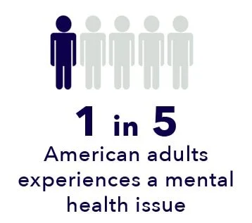
Note: The content on Sleepopolis is meant to be informative in nature, but it shouldn’t take the place of medical advice and supervision from a trained professional. If you feel you may be suffering from any sleep disorder or medical condition, please see your healthcare provider immediately.
Mental Illness: Causes and Symptoms
Mental health problems can affect self-esteem, happiness, and basic functions, such as eating and sleeping. (1) Social and financial success is strongly influenced by psychological wellness, including self-awareness and our ability to exercise self-control.
Many factors contribute to mental health issues, including:
Genes and brain chemistry
Family history of mental health problems
Life experiences, such as trauma or abuse
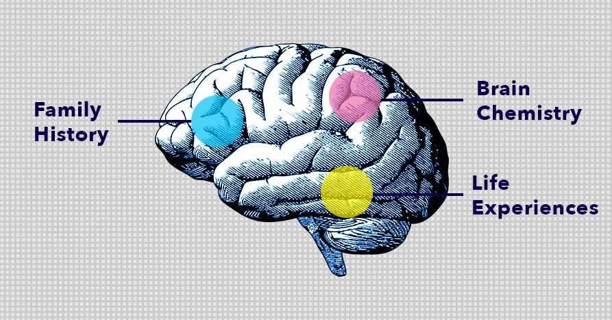
Mental health issues run the gamut from minor difficulty with daily living to full-blown psychosis. Treatment can have a significant impact on the course and outlook of psychological issues, making early detection crucial.
Mental illness can appear in early childhood or not until adulthood. (2) Some psychological issues may not emerge until later in life and can be influenced by certain drugs and medical conditions. There are numerous warning signs of mental health issues. These vary depending on the nature of the condition, and may include:
Eating or sleeping too much or too little
Withdrawing from relationships and usual activities
Having low or no energy
Feeling numb
Experiencing unexplained aches and pains
Feeling helpless or hopeless
FAQ
Q: What is the most common mental illness?A: Depression, a mood disorder that affects 300 million people worldwide, is the single most common mental illness. Anxiety disorders are more prevalent overall.
Psychological difficulties increase the risk of troubled relationships with family and friends. (3) Interpersonal conflict can be caused by irritability and mood swings, or feelings such as fear and anger. Other cognitive signs associated with mental health issues include worry, confusion, memory loss, and intrusive thoughts.
More serious mental disorders may cause hallucinations, including hearing voices or believing things that are not true. Suicidal behavior or thoughts of harming another person are also common signs of psychological issues. A person suffering from mental illness may experience paranoia, imagined physical ailments, or the need to abuse drugs or alcohol.
Types of Mental Health Disorders
Each type of mood disorder is characterized by unique signs and symptoms and may co-occur with other disorders. For example, anxiety might occur along with substance abuse, physical complaints, or a sleep disorder like insomnia.
The following are some of the most common mental health disorders:
Anxiety Disorders. People with anxiety disorders respond to certain objects or situations with fear and dread. Anxiety disorders can include panic disorders, generalized anxiety disorder, and phobias. (4) Symptoms of anxiety include:
Fatigue
Feelings of restlessness or irritability
Difficulty focusing or learning new concepts
Chronic pain or muscle tension
Trouble controlling feelings of worry
Problems sleeping, including the inability to sleep, unsatisfying sleep, or excessive sleeping
Psychiatric Disorders in Children. Common psychiatric disorders in children include attention deficit hyperactivity disorder (ADHD) a neurodevelopmental disorder that usually persists into adulthood. Symptoms include impulsiveness, trouble focusing on tasks or conversations, restlessness, and feeling easily frustrated. Oppositional defiant disorder is part of a spectrum known as disruptive, impulse-control, and conduct disorders. Symptoms of oppositional defiant disorder include hostility toward and lack of cooperation with teachers, parents, and other authority figures. (5)
Eating Disorders. Eating disorders involve extreme emotions, attitudes, and behaviors related to weight and food. Common eating disorders include anorexia, bulimia, and binge eating.
Substance Use Disorders. Mental health problems and substance abuse disorders often occur together. (6) Substance abuse can be a cause or a result of psychiatric issues, and may be an attempt by some patients to self-medicate or reduce symptoms.
Mood Disorders. Almost one in ten people aged eighteen or older have a mood disorder. Mood disorders involve persistent feelings of sadness, or feelings that fluctuate between extreme happiness and extreme sadness. Mood disorders include:
Depression. Approximately 17.3 million people in the US — 7.1% of adults — suffered from one or more major depressive episodes in the last year. Depression is characterized by sadness and lack of usual enjoyment that continue for two weeks or longer. Grief and sadness related to a change in life circumstances are different from depression, which doesn’t improve in response to external events
Bipolar disorder. Bipolar disorder affects approximately six million adults in the US, and impacts men and women equally. The disorder is characterized by severe fluctuations in mood, behavior, and thought patterns. Fluctuations can last from a few hours to several months, and may be affected seasonal and light changes
Personality Disorders. People with personality disorders have inflexible personality traits that are distressing to the sufferer and may cause problems in work, school, or social relationships. (7) These disorders are highly resistant to treatment, and include the following:
Antisocial personality disorder, or APD. People with antisocial personality disorder exploit, manipulate, and disregard the feelings of others. APD describes the behavior of sociopaths and psychopaths. Psychopaths do not have a conscience, whereas sociopaths have a conscience that is severely disordered
Narcissistic personality disorder, or NPD. Approximately 1% of people are afflicted with NPD. The disorder is associated with lack of empathy, feelings of superiority, and a need for admiration. Narcissists tend to be easily hurt and are unable to tolerate criticism. Treatment is generally ineffective because people with NPD don’t believe they have a mental health issue
Borderline personality disorder, or BPD. People with borderline personality disorder have trouble regulating their emotions. They tend to act impulsively, resulting in career and relationship difficulties. Of the 1.4% of the population with BPD, 75% are women, though this number may represent misdiagnosis in many men with the disorder
Psychotic Disorders. Psychotic disorders are characterized by abnormal perceptions such as hallucinations and delusions. Hallucinations are false perceptions that involve hearing or seeing things that don’t exist. Delusions are false beliefs, such as being pursued by authorities or people wishing to do the sufferer harm. The most common psychotic disorder is schizophrenia, which typically appears in the mid to late twenties. Possible causes of schizophrenia include genetic and environmental factors, disordered brain circuitry, trauma, and drug abuse.
Psychosis
Psychosis describes mental conditions that involve loss of contact with reality.
Mental Health and Sleep
One common sign of mental health difficulty is trouble sleeping. (8) The sleep disorder most closely associated with psychiatric disturbances is chronic insomnia. Some common mental illnesses and their effect on sleep include the following:
Depression. Clinical depression often presents with persistent insomnia, or the inability to sleep. (9) Hypersomnia, or excessive drowsiness, can be a sign of depression, as well. Sleep disorders are associated so strongly with depression that some medical practitioners advise caution in diagnosing depression in patients without symptoms of a sleep disorder. Up to 75% of people diagnosed with depression also suffer from insomnia.
Bipolar Disorder. Bipolar disorder is strongly associated with the inability to fall asleep, stay asleep, fall back to sleep, or all three. People in the manic phase of bipolar disorder often have difficulty sleeping, whereas those in the depressive phase may sleep much more than usual. Studies show that sleep deprivation can aggravate the struggles with emotional regulation that are common in people who are bipolar. (10) For many sufferers, trouble sleeping can be an indication of an approaching manic phase.
Anxiety Disorder. Anxiety is a principal cause of chronic insomnia. Even common anxiety related to work and life circumstances may trigger insomnia. A primary cause of chronic insomnia is conditioned anxiety related to sleep and the bedtime routine. Anxiety’s impact on the body includes hyperactivity of the central nervous system and excessive release of stress hormones, such as adrenaline and cortisol. Studies show a strong association between a history of insomnia and anxiety disorder, panic disorder, and social anxiety disorder. (11)
Borderline Personality Disorder. BPD sufferers with insomnia typically experience the daytime consequences of sleep loss, including sleepiness, difficulty concentrating, and irritability. Symptoms of BPD can be worsened by insomnia, leading to a vicious cycle of sleeplessness and behavioral issues.
Personality Disorders. Some personality disorders may be associated with disturbed sleep patterns. (12) One study revealed that people with circadian rhythm disorder are more likely to have a personality disorder than people with normal sleep and wake cycles.
Psychotic Disorders. Psychotic disorders such as schizophrenia can have a profound impact on sleep. Sleep disturbances are often an early sign of schizophrenia and can precede psychiatric symptoms by months or years. Schizophrenia patients are more likely to suffer from other sleep disorders, as well, including:
Obstructive sleep apnea
Restless legs syndrome
Periodic limb movement disorder
Circadian rhythm dysfunction
FAQ
Q: What is circadian rhythm dysfunction?A: Circadian rhythm dysfunction is caused by a loss of synchronization between sleep-wake cycles and natural light and darkness signals. Symptoms include the inability to fall or stay asleep, cognitive dysfunction, and trouble maintaining a traditional sleep schedule.
Does Depression Cause Insomnia, or Vice Versa?
It was once thought that insomnia symptoms resulted from psychiatric disorders and depression, not the other way around. Now the evidence isn’t so clear. Sleep problems may not just be the result of emotional disturbances, they may increase the likelihood of suffering from them, as well.
Medications to treat psychiatric disturbances may also cause insomnia. Psychotropic medications can have stimulating effects that contribute to interrupted sleep. Restless leg syndrome and periodic limb movements may be triggered or exacerbated by antidepressants and other drugs used to treat mood disorders.
An analysis of major studies revealed that volunteers who suffered from insomnia symptoms were twice as likely to be diagnosed with depression as those without sleep difficulties. Why? Though the connection between mood disorders and insomnia isn’t entirely understood, it is well-known that hormones and neurotransmitters are affected by poor sleep. Sleep is a restorative activity that reduces stress in the body and areas of the brain, especially the axis between the hypothalamus and adrenal and pituitary glands. Excess secretion of cortisol — also known as “the stress hormone” — may play a role, as well.
Lack of sleep and the resulting physiological stress may predispose insomnia sufferers to major mood disturbances, making early treatment of sleep disorders essential, especially in people with a family history of mental health issues. (13)
Depression and Other Sleep Disorders
Insomnia is not the only sleep disorder associated with depression and anxiety. (14) Sleep apnea is a common cause of depression, as is hypersomnia. Sleep apnea is characterized by blockage of the airway and repeated awakenings during the night. Chronic sleep apnea can result in weight gain, increased risk of heart attack and stroke, and memory problems.
Hypersomnia is associated with excessive time spent sleeping and daytime sleepiness. The disorder can be caused by medical conditions, certain drugs, and immune system dysfunction. Though most people need between seven and nine hours of sleep each night, the need for significantly more can indicate a mood disorder. Like sleep apnea, hypersomnia is related to a greater likelihood of heart attack and stroke.
Hypersomnia
A sleep disorder associated with excessive sleeping. Hypersomnia includes narcolepsy, an autoimmune disease that can cause extreme sleepiness and muscle weakness.
Psychiatric Medications and Sleep
Medications to treat psychological disturbances can have a positive or negative impact on sleep. (15) Most antidepressant medications influence the neurotransmitters dopamine, serotonin, and norepinephrine, all of which help to regulate sleep and wake cycles. Some can have stimulating effects that contribute to insomnia.
Restless legs syndrome and periodic limb movements can be triggered or exacerbated by antidepressants and other drugs used to treat mood disorders. (16) These medications can be helpful in patients without movement-related sleep disorders who suffer from hypersomnia.
Other medications such as older tricyclic drugs can help establish healthy sleep patterns in depressed patients with insomnia. Once a depressed patient starts taking medication, insomnia may be the last symptom to improve. Newer antidepressants such as selective serotonin reuptake inhibitors can suppress the REM stage of sleep, as well as the vivid dreaming that occurs during REM sleep.
Antipsychotic medications can help with insomnia, a common issue for schizophrenia sufferers. Some of these drugs can also cause daytime sleepiness, which may be preferable to insomnia. (17) Hypnotic medications are often prescribed along with antipsychotics, and may initially help with insomnia due to their sedative effects. However, many patients become tolerant of hypnotics or develop a rebound response, which can limit their usefulness for insomnia over the long-term.
Suicidal Behavior and Sleep
Suicidal behavior is a common feature of certain psychiatric conditions, including bipolar disorder and depression. Suicidal thoughts and actions can also occur in the absence of a psychiatric condition. Suicide is the second leading cause of death in people between 15 and 24. Suicide is more likely to be completed by people with access to guns.
Signs of suicidal thoughts and behavior may include the following:
Speaking about suicide or the desire to die
Feeling or talking about feeling hopeless
Researching ways to commit suicide
Appearing anxious or depressed
Excessive use of alcohol, or use of drugs
Exhibiting unusual behavior, such as rage, mood swings, or agitation
Suicidal behavior is often caused by psychiatric conditions, but may also be triggered by difficult life events, stress, loss of a loved one, or past trauma such as emotional, physical, or sexual abuse. Suicidal behavior is also more common in people suffering from post-traumatic stress disorder or chronic pain.
Recent research reveals that suicidal behavior and lack of sufficient sleep are related in adolescents and adults. (18) People who completed their suicidal actions were more likely to have suffered from insomnia, hypersomnia, or another sleep disturbance. Though further research is needed to better understand this link, existing studies reveal a strong association between mental health and sleep.
FAQ
Q: What is suicidal ideation?A: Suicidal ideation involves thinking about, planning, or considering suicide.
Last Word From Sleepopolis
Mental health is critically important to happiness and well-being. Research reveals a connection between mental illness and disturbed sleep, an association that may exist months or years before psychiatric symptoms appear.
Medications and other mental health treatments can help regulate sleep patterns, but may also cause or worsen insomnia, hypersomnia, and other sleep difficulties. Adjustment of medications or separate treatment of sleep issues can help establish healthy sleep patterns and improve quality of life for people suffering from mental health disorders.
References
Amy C. Watson, Self-Stigma in People With Mental Illness, Schizophrenia Bulletin, January 25, 2007
William Copeland Ph.D., Cumulative Prevalence of Psychiatric Disorders by Young Adulthood: A Prospective Cohort Analysis From the Great Smoky Mountains Study, Journal of the American Academy of Child & Adolescent Psychiatry, March 2011
Alan R. Teo, Social Relationships and Depression: Ten-Year Follow-Up from a Nationally Representative Study, Plos One, April 30, 2013
Peter J. Norton, Transdiagnostic models of anxiety disorder: Theoretical and empirical underpinnings, Clinical Psychology Review, August 2017
Martin B. Keller, MD, The Disruptive Behavioral Disorder in Children and Adolescents: Comorbidity and Clinical Course, Journal of the American Academy of Child & Adolescent Psychiatry, March 1992
Robert E Drake, et al. A systematic review of psychosocial research on psychosocial interventions for people with co-occurring severe mental and substance use disorders, Journal of Substance Abuse Treatment, January 2008
Tyrer P, Mulder R, Crawford M, Newton-Howes G, Simonsen E, Ndetei D, Koldobsky N, Fossati A, Mbatia J, Barrett B., Personality disorder: a new global perspective, World Psychiatry, February 2010
Krahn LE., Psychiatric disorders associated with disturbed sleep, Seminars in Neurology, March 25, 2005
Chiara Baglioni et al., Insomnia as a predictor of depression: A meta-analytic evaluation of longitudinal epidemiological studies, Journal of Affective Disorders, December 2011
Harvey AG, Talbot LS, Gershon A, Sleep Disturbance in Bipolar Disorder Across the Lifespan, Clinical Psychology, New York State Psychiatric Institute, April 8, 2012
Heidemarie Blumenthal, et al., The Links Between Social Anxiety Disorder, Insomnia Symptoms, and Alcohol Use Disorders: Findings From a Large Sample of Adolescents in the United States, Behavior Therapy, January 2019
Yaron Dagan, High prevalence of personality disorders among Circadian Rhythm Sleep Disorders (CRSD) patients, Journal of Psychosomatic Research, October 1996
Liu X, Buysse DJ, Gentzler AL, Kiss E, Mayer L, Kapornai K, Vetró A, Kovacs M., Insomnia and hypersomnia associated with depressive phenomenology and comorbidity in childhood depression, Sleep, January 30, 2007
Nutt D, Wilson S, Paterson L., Sleep disorders as core symptoms of depression, Dialogues in Clinical Neuroscience, September 2008
DeMartinis NA, Winokur A., Effects of psychiatric medications on sleep and sleep disorders, CNS and Neurological Disorders Drug Targets, February 6, 2007
Staner L., Sleep disturbances, psychiatric disorders, and psychotropic drugs, Dialogues in Clincal Neuroscience, December 7, 2005
Waite F, Myers E, Harvey AG, Espie CA, Startup H, Sheaves B, Freeman D., Treating Sleep Problems in Patients with Schizophrenia, Behavioural and Cognitive Psychotherapy, May 2016
Goldstein TR, Bridge JA, Brent DA., Sleep Disturbance Preceding Completed Suicide in Adolescents, Journal of Consulting and Clinical Psychology, February 17, 2010
Original article: https://sleepopolis.com/education/how-does-mental-health-affect-your-sleep/
Rose MacDowell
Rose is the Chief Research Officer at Sleepopolis, which allows her to indulge her twin passions for dense scientific studies and writing about health and wellness. An incurable night owl, she loves discovering the latest information about sleep and how to get (lots) more of it. She is a published novelist who has written everything from an article about cheese factories to clock-in instructions for assembly line workers in Belgium. One of her favorite parts of her job is connecting with the best sleep experts in the industry and utilizing their wealth of knowledge in the pieces she writes. She enjoys creating engaging articles that make a difference in people’s lives. Her writing has been reviewed by The Boston Globe, Cosmopolitan, and the Associated Press, and received a starred review in Publishers Weekly. When she isn’t musing about sleep, she’s usually at the gym, eating extremely spicy food, or wishing she were snowboarding in her native Colorado. Active though she is, she considers staying in bed until noon on Sundays to be important research.

How caloric restriction prevents negative effects of aging in cells
Peas on plate, dieting concept (stock image). Credit: © Studio KIVI / Adobe Stock
How caloric restriction prevents negative effects of aging in cells
February 27, 2020
Science Daily/Salk Institute
A new study provides the most detailed report to date of the cellular effects of a calorie-restricted diet in rats. While the benefits of caloric restriction have long been known, the new results show how this restriction can protect against aging in cellular pathways.
If you want to reduce levels of inflammation throughout your body, delay the onset of age-related diseases, and live longer, eat less food. That's the conclusion of a new study by scientists from the US and China that provides the most detailed report to date of the cellular effects of a calorie-restricted diet in rats. While the benefits of caloric restriction have long been known, the new results show how this restriction can protect against aging in cellular pathways, as detailed in Cell on February 27, 2020.
"We already knew that calorie restriction increases life span, but now we've shown all the changes that occur at a single-cell level to cause that," says Juan Carlos Izpisua Belmonte, a senior author of the new paper, professor in Salk's Gene Expression Laboratory and holder of the Roger Guillemin Chair. "This gives us targets that we may eventually be able to act on with drugs to treat aging in humans."
Aging is the highest risk factor for many human diseases, including cancer, dementia, diabetes and metabolic syndrome. Caloric restriction has been shown in animal models to be one of the most effective interventions against these age-related diseases. And although researchers know that individual cells undergo many changes as an organism ages, they have not known how caloric restriction might influence these changes.
In the new paper, Belmonte and his collaborators -- including three alumni of his Salk lab who are now professors running their own research programs in China -- compared rats who ate 30 percent fewer calories with rats on normal diets. The animals' diets were controlled from age 18 months through 27 months. (In humans, this would be roughly equivalent to someone following a calorie-restricted diet from age 50 through 70.)
At both the start and the conclusion of the diet, Belmonte's team isolated and analyzed a total of 168,703 cells from 40 cell types in the 56 rats. The cells came from fat tissues, liver, kidney, aorta, skin, bone marrow, brain and muscle. In each isolated cell, the researchers used single-cell genetic-sequencing technology to measure the activity levels of genes. They also looked at the overall composition of cell types within any given tissue. Then, they compared old and young mice on each diet.
Many of the changes that occurred as rats on the normal diet grew older didn't occur in rats on a restricted diet; even in old age, many of the tissues and cells of animals on the diet closely resembled those of young rats. Overall, 57 percent of the age-related changes in cell composition seen in the tissues of rats on a normal diet were not present in the rats on the calorie restricted diet.
"This approach not only told us the effect of calorie restriction on these cell types, but also provided the most complete and detailed study of what happens at a single-cell level during aging," says co-corresponding author Guang-Hui Liu, a professor at the Chinese Academy of Sciences.
Some of the cells and genes most affected by the diet related to immunity, inflammation and lipid metabolism. The number of immune cells in nearly every tissue studied dramatically increased as control rats aged but was not affected by age in rats with restricted calories. In brown adipose tissue -- one type of fat tissue -- a calorie-restricted diet reverted the expression levels of many anti-inflammatory genes to those seen in young animals.
"The primary discovery in the current study is that the increase in the inflammatory response during aging could be systematically repressed by caloric restriction" says co-corresponding author Jing Qu, also a professor at the Chinese Academy of Sciences.
When the researchers homed in on transcription factors -- essentially master switches that can broadly alter the activity of many other genes -- that were altered by caloric restriction, one stood out. Levels of the transcription factor Ybx1 were altered by the diet in 23 different cell types. The scientists believe Ybx1 may be an age-related transcription factor and are planning more research into its effects.
"People say that 'you are what you eat,' and we're finding that to be true in lots of ways," says Concepcion Rodriguez Esteban, another of the paper's authors and a staff researcher at Salk. "The state of your cells as you age clearly depends on your interactions with your environment, which includes what and how much you eat."
The team is now trying to utilize this information in an effort to discover aging drug targets and implement strategies towards increasing life and health span.
https://www.sciencedaily.com/releases/2020/02/200227144259.htm
Drinks with added sugars linked to lipid imbalance, which increases CVD risk
February 26, 2020
Science Daily/American Heart Association
Drinking 12 ounces of sugary drinks more than once per day is linked to lower levels of high-density lipoprotein cholesterol (HDL-C), ("good" cholesterol), and higher levels of triglycerides, in middle aged and older adults, both of which have been shown to increase risk of cardiovascular disease. These results are from a new observational study published today in the Journal of the American Heart Association, the open access journal of the American Heart Association.
In previous studies, added sugars have been shown to increase cardiovascular disease risk. Beverages such as sodas, sports drinks and fruit-flavored drinks are the largest source of added sugars for Americans.
"For some time, we have known sugary drinks can have a negative effect on Americans' health status, yet the assumption for many is that they only contribute to weight gain," said Eduardo Sanchez, M.D., M.P.H., chief medical officer for prevention and chief of the Center for Health Metrics and Evaluation at the American Heart Association. "This research reinforces our understanding of the potential negative impact sugary drinks have on blood cholesterol, which increases heart disease risk. It is yet one more reason for all of us to cut back on sodas and other sugar-sweetened beverages."
Researchers hypothesized that dyslipidemia could be one pathway by which sugary drinks may increase cardiovascular disease risk. An estimated 40% to 50% of U.S. adults are affected by dyslipidemia, an unhealthy imbalance of cholesterol and triglyceride levels in the blood, which increases the risk of cardiovascular disease.
To determine the association between sugary drinks on triglyceride and cholesterol levels, researchers studied observational medical data of up to 5,924 participants from the Offspring and Generation 3 cohorts of the Framingham Heart Study, who were followed for an average of 12.5 years between 1991 and 2014. The Offspring cohort of the Framingham Heart Study includes the children of original participants in the Framingham Heart Study, and the Generation 3 cohort includes grandchildren of the original participants in the Framingham Heart Study.
For this study, the beverages were defined as: 12 ounces of sugary drinks, such as sodas, fruit-flavored drinks, sports drinks, presweetened coffees and teas; 12 ounces of low-calorie sweetened beverages, including naturally and artificially sweetened "diet" sodas or other flavored drinks; or 8 ounces of 100% fruit juices, including orange, apple, grapefruit and other juices derived from whole fruits with no added sugars. Study participants were classified into five groups according to how often they drank the different beverage types ranging from low intake (<1 serving per month) to high intake (>1 serving per day).
Researchers analyzed how the different drink types and their consumption levels correlated with changes in cholesterol and triglyceride levels over approximately four-year periods. They found that:
Drinking sugar-sweetened beverages (more than 12 ounces per day) was associated with a 53% higher incidence of high triglycerides and a 98% higher incidence of low HDL cholesterol (the "good" cholesterol) compared to those who drank less than one serving per month;
Drinking low-calorie sweetened beverages did not appear to be associated with increased dyslipidemia risk among the people who regularly drank low-calorie sweetened beverages; and
Regularly drinking up to 12 ounces of 100% fruit juice per day was not associated with adverse changes in cholesterol or dyslipidemia, though further research is needed to warrant this finding.
"Reducing the number of or eliminating sugary drink consumption may be one strategy that could help people keep their triglyceride and HDL cholesterol at healthier levels," said lead study author Nicola McKeown, Ph.D., a nutrition epidemiologist at the Jean Mayer USDA Human Nutrition Research Center on Aging at Tufts University in Boston. "And, while our study didn't find negative consequences on blood lipids from drinking low-calorie sweetened drinks, there may be health consequences of consuming these beverages on other risk factors. Water remains the preferred and healthiest beverage."
While previous cross-sectional studies have had similar findings, this study reaffirms those findings with prospective data. One potential limitation of the study is that participants self-reported their dietary intake.
The American Heart Association recommends people eliminate sugary drink consumption to improve heart health and to reduce the risk of cardiovascular disease.
The National Institutes of Health, the American Heart Association and the U.S. Department of Agriculture's Agricultural Research Service supported the study.
https://www.sciencedaily.com/releases/2020/02/200226072110.htm